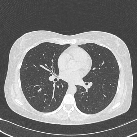
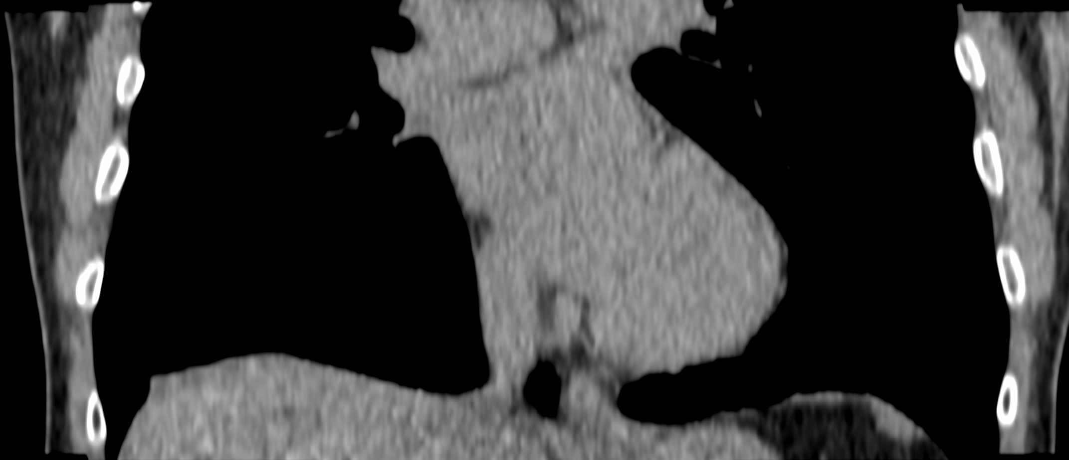
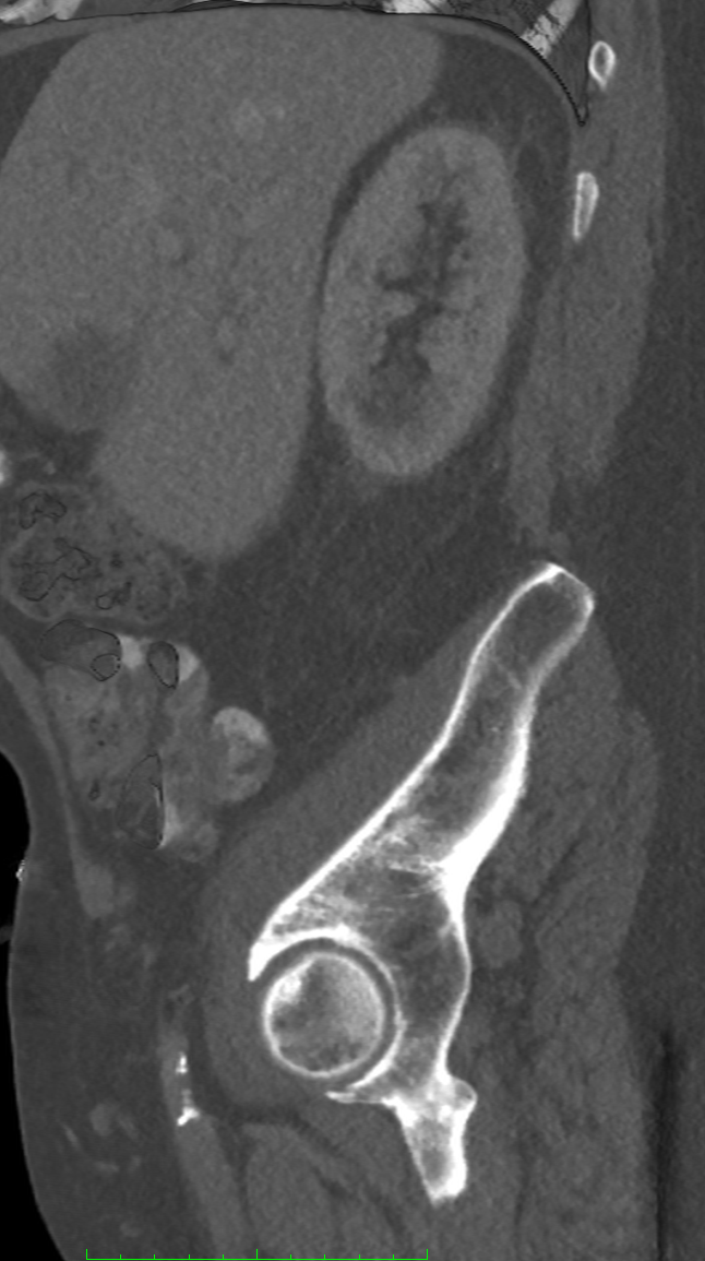
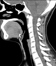
Vertebrae or hip detected; vertebrae segmented, measured, fracture detection
Automatically detects vertebrae or hip present on the scan
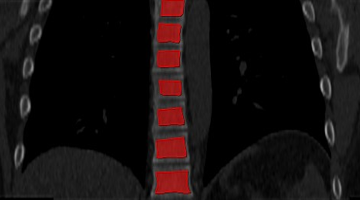
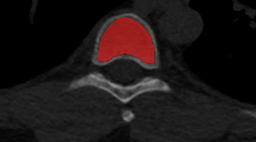
Segments individual vertebrae
Measures vertebral height
Identifies fractures
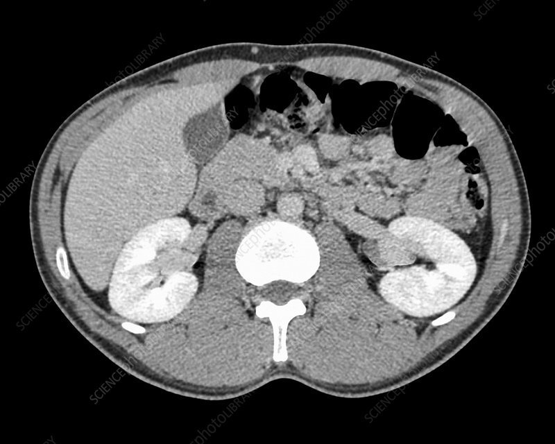
Step 2
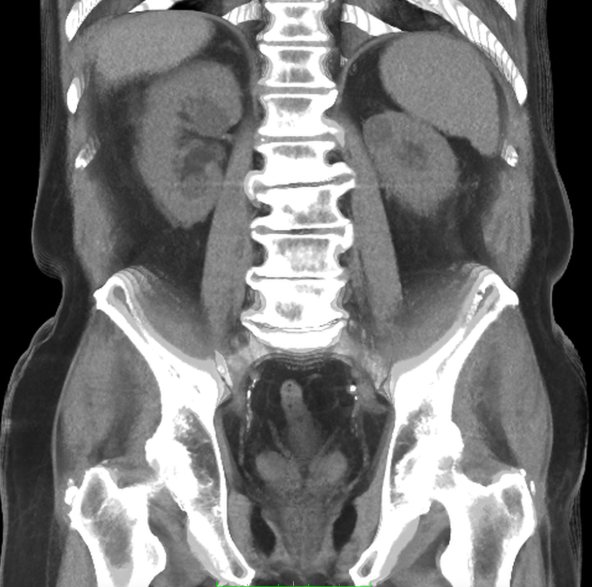
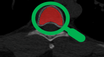
Trabecular part of vertebra or hip detected
Detects the trabecular component of the vertebral or hip bones, excluding cortical bone
Step 3
Measures trabecular bone for each; determines bone density
Accurately measures and averages the Hounsfield units of trabecular bone within each vertebrae and hip, avoiding fractures.
Then puts into formula (BDI patented) to convert to bone density – adjusted for CT scanner, spinal and hip location.
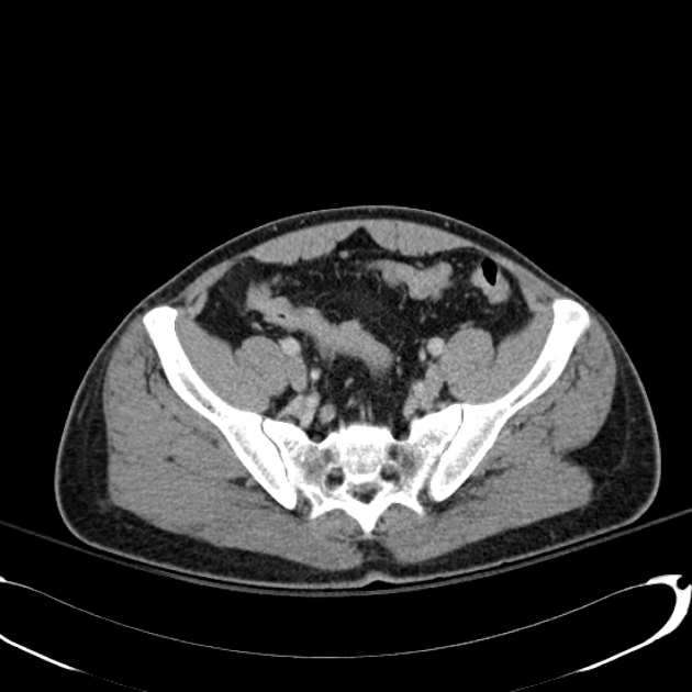
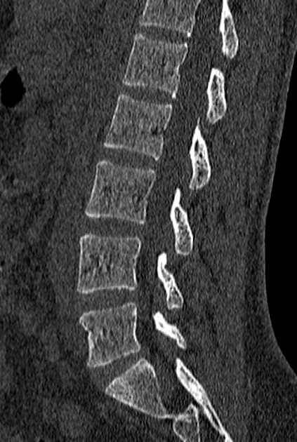
Step 4
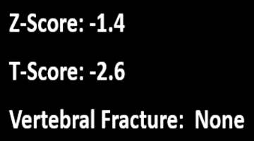
Calculates/reports T-Score, Z-Score, vertebral fracture presence and severity
Calculates & reports T-Score and Z-Score (gold standard metrics for osteoporosis analysis) by comparing bone density values to age , gender and race/ethnicity population assessments
Determines/reports any vertebral fracture presence and severity
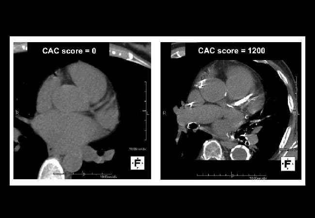
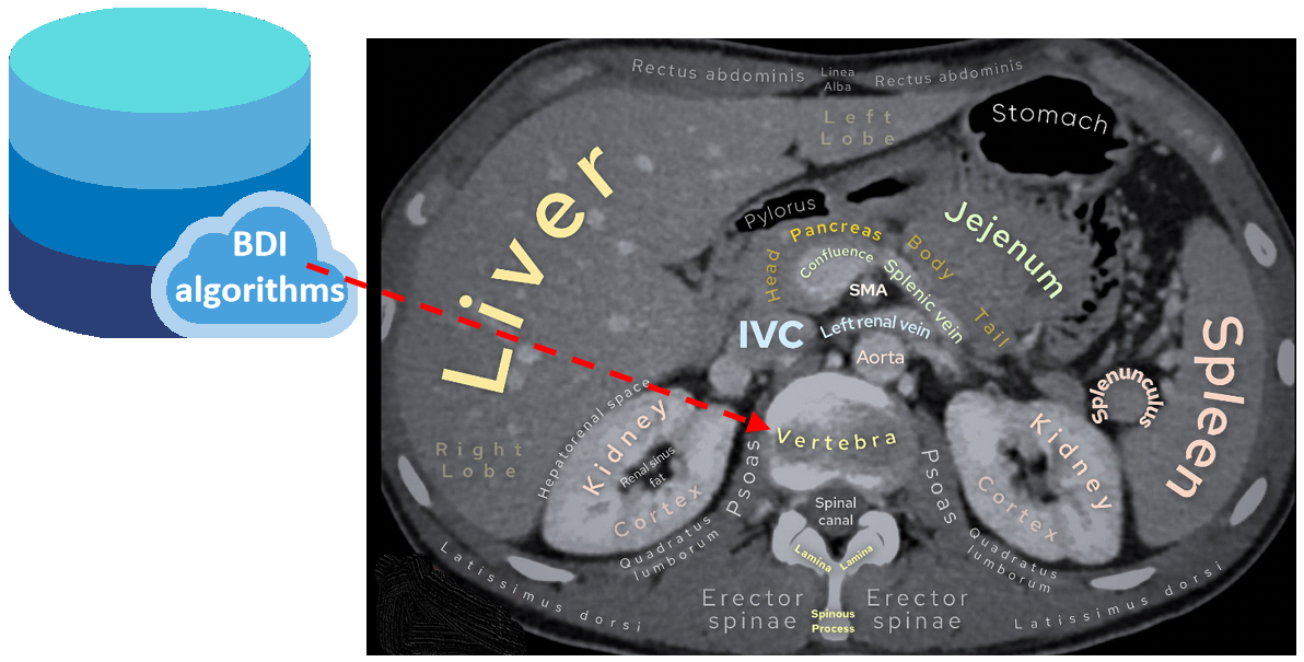
We utilize the subject’s heart density, chest wall/abdominal or hip fat to calibrate the patient, CT scanner, and scan parameters to ensure the most accurate measure of individual BMD
Step 5
BDI expert physician reviews and finalizes report
Expert BDI Physician reviews, modifies if necessary, and finalizes report
Step 6
BDI Report
BDI final report in natural language
Sent to provider who ordered BDI osteoporosis screening and others as desired
Contact us to learn more about BDI’s osteoporosis screening service